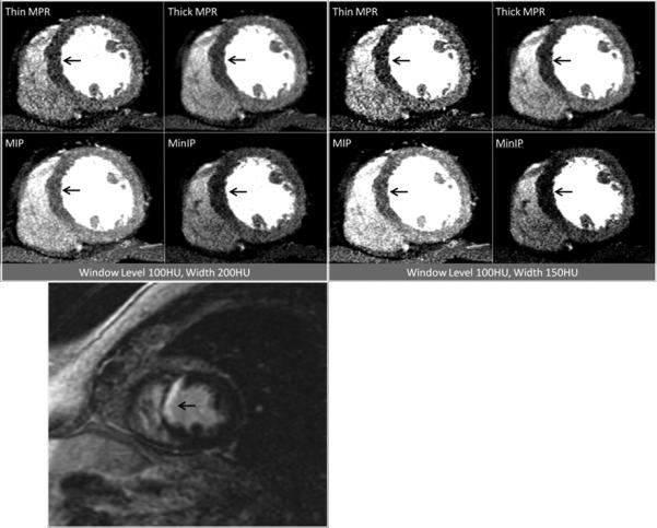
Figure 1.

Contrast-enhanced short axis CT images at window level 100HU and width 200HU as well as window level 100HU and width 150HU demonstrating anteroseptal and inferoseptal myocardial infarction at the mid ventricular level (arrows), as visualized by the four post-processing techniques. The patient was a 63 year-old man with myocardial infarction. Corresponding delayed enhancement short axis MRI image are presented for correlation (bottom).
