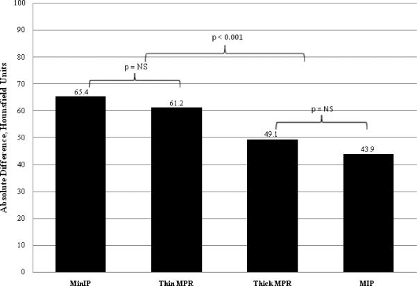
Figure 4.

Average absolute difference in Hounsfield Unit attenuation between areas of infarcted myocardium and areas of remote normal myocardium. Segments processed with the MinIP and thin MPR techniques had a significantly greater difference than segments processed with the thick MPR and MIP techniques. There was no significant difference between segments processed with the MinIP and thin MPR techniques nor between segments processed with the thick MPR and MIP techniques.
