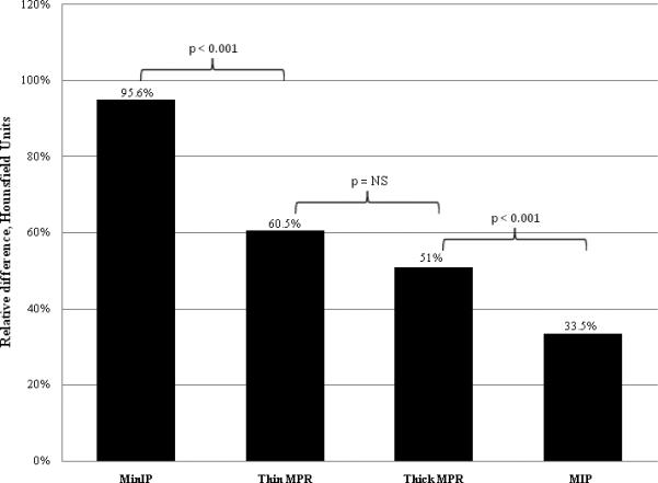
Figure 5.

Average relative difference in Hounsfield Unit attenuation between areas of infarcted myocardium and areas of remote normal myocardium. Segments processed with the MinIP technique had a significantly greater relative difference than segments processed with thin MPR or thick MPR, which in turn had a significantly greater difference than segments processed with the MIP techniques. There was no significant difference between segments processed with the thin MPR and thick MPR techniques.
