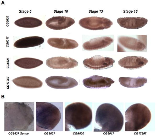
Figure 3. Tissue-specific enrichment of palmitoylome genes.

A. This figure catalogs distinct tissue-specific patterns of expression for several of the DHHC proteins as determined by in situ hybridization on mixed-stage Drosophila embryos. The anti-sense staining for four different DHHC genes at four stages of embryonic development is shown. Lateral views of a developmental expression series for each gene proceed from left to right across the figure. A ventral view is shown for the stage 16 CG5620 in situ. Anterior is to the right. Only the DHHC genes that showed obvious enrichment in particular tissues are presented. The rest of the in situ hybridization results are shown in Table 3. B. The anti-sense staining of 3rd instar larval brains for four different DHHC genes is shown. Each image is a close-up of one brain lobe. An example of a sense control for CG6627 is also shown.
