
Figure 9. CG18815 thioesterase is the putative Drosophila APT1 ortholog.
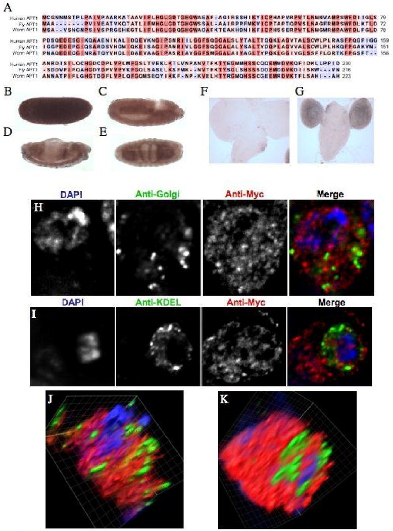
A. Sequence alignment of the human, Drosophila, C. elegans APT1 amino acid sequence generated with ClustalX. The amino acid residue's background color indicates the degree of conservation between the proteins: red is highly conserved and blue indicates no conservation. B-E. The anti-sense staining for CG18815 at four stages of embryonic development is shown. B. Stage 5. C. Stage 10. D. Stage 13. E. Stage 16. Lateral views of a developmental expression series are shown except for the ventral view that is shown for the stage 16 CG18815 in situ. Anterior is to the right. F. A sense probe image of a 3rd instar larval brain CG18815 in situ. G. An image showing specific brain lobe staining in 3rd instar larvae for the CG18815 anti-sense probe. H. Images of fixed S2 cells transiently transfected with a CG18815-6×Myc fusion protein and co-stained with DAPI and an anti-Golgi antibody. I. Images of fixed S2 cells transiently transfected with a CG18815-Myc fusion protein and co-stained with DAPI and an anti-KDEL antibody to mark the ER. J. A volume view produced from a deconvolved Z-stack of the cell shown in C. K. A volume view produced from a deconvolved Z-stack of the cell shown in D.