Fig. 1.
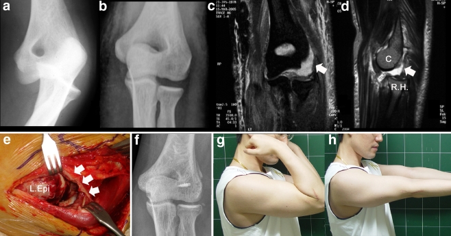
Case 4: a 27-year-old man with injuries of the lateral collateral ligament and common extensor tendon. a Preoperative plain radiograph. b Post-reduction radiograph. c, d Post-reduction magnetic resonance image showing injuries of the lateral collateral ligament complex (white arrows) with posterolateral subluxation of the radial head (R.H.) from the capitellum (C). e Intraoperative photograph of the lateral epicondyle (L. Epi). Note the complete avulsion of the collateral ligament, common extensor tendon from the lateral epicondyle. f Postoperative radiograph. g, h Photographs at last follow-up

