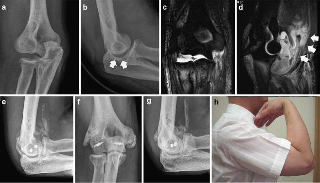
Fig. 3.
Case 12: a 40-year-old man with injuries of both medial and lateral structures. a Initial plain radiograph. b Non-congruent ulnohumeral joint after closed reduction (white arrows). c, d Post-reduction magnetic resonance image showing injuries of the medial, lateral collateral ligament complex and anterior capsule with large haematoma (white arrows) in the anterior compartment. e Calcification in the brachialis which limited range of motion during rehabilitation. f, g Plain radiographs at 32 months postoperatively. h Photograph at last follow-up

