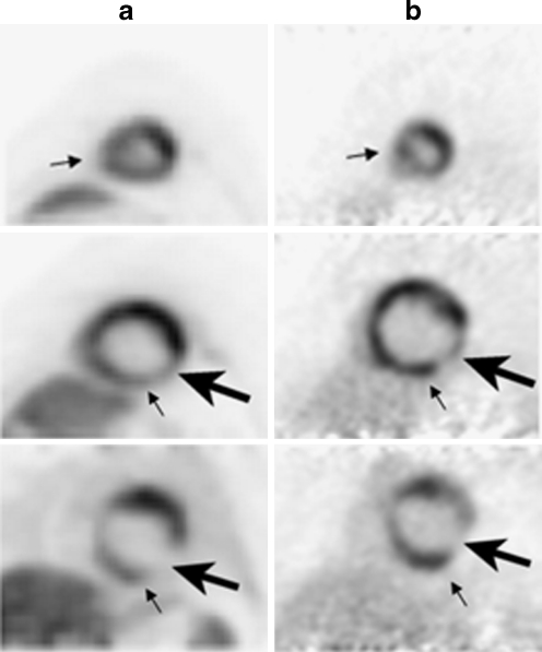
Fig. 1.
13N-ammonia axial PET myocardial perfusion images (column A) and 18F-FDG axial PET myocardial metabolism images (column B) in a 49-year-old male history of cardiomyopathy. Images demonstrate a mismatched pattern of decreased myocardial perfusion with preserved 18F-FDG metabolism (small arrows) in the inferoseptal and inferior walls, suggesting the presence of viable hibernating myocardium. Furthermore, there is a matched pattern of decreased myocardial perfusion and metabolism (large arrow) in the proximal to mid inferolateral wall, which is compatible with an infarct

