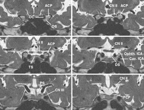
Fig. 2.
Sequential T2-weighted three-dimensional fast spin-echo magnetic resonance imaging in oblique coronal planes, photographed from anterior to posterior, shows the ICA in the paraclinoid region and its surrounding anatomic structures. ACP : anterior clinoid process, Cav. : cavernous, CN II : optic nerve, CN III : oculomotor nerve, DS : diaphragma sellae, ICA : internal carotid artery, OC : optic canal, OS : optic strut, Ophth. : ophthalmic, PCP : posterior clinoid process, PS : pituitary stalk, TS : tuberculum sellae.

