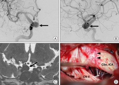
Fig. 3.
A 48-year-old man was incidentally diagnosed to have a intracranial aneurysm at the paraclinoid ICA. A and B : Preoperative anteroposterior (A) and lateral (B) angiogram show a large aneurysm (arrow) involving the right paraclinoid ICA at the level of the ophthalmic artery origin. It would be difficult to localize the aneurysm in relation to the distal dural ring, by using the level of ophthalmic artery origin as a landmark. C : T2-weighted three-dimensional fast spin-echo magnetic resonance imaging in the oblique coronal plane shows an aneurysm (arrow) within the cistern, arising from the medial aspect of the intradural ICA. D : Intraoperative photograph shows a superolateral view of the right paraclinoid region. The anterior clinoid process was drilled, exposing the aneurysm neck (arrow) protruding medially above the dissected distal dural ring (arrowheads). Clin. : clinoid, CN II : optic nerve, ICA : internal carotid artery.

