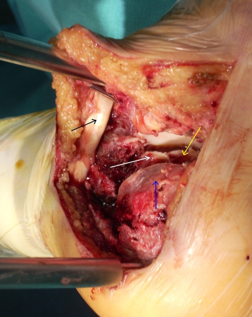
Fig. 3.
Calcaneal fracture, Sanders type III. PTC distraction with the use of LBD. Black arrow points out the peroneal tendons. Deep inside the joint, a yellow arrow points to the undislocated anteromedial articular fragment. The mid portion of PAS is marked with a white arrow. The depressed and anteriorly rotated lateral fragment is shown by a blue arrow

