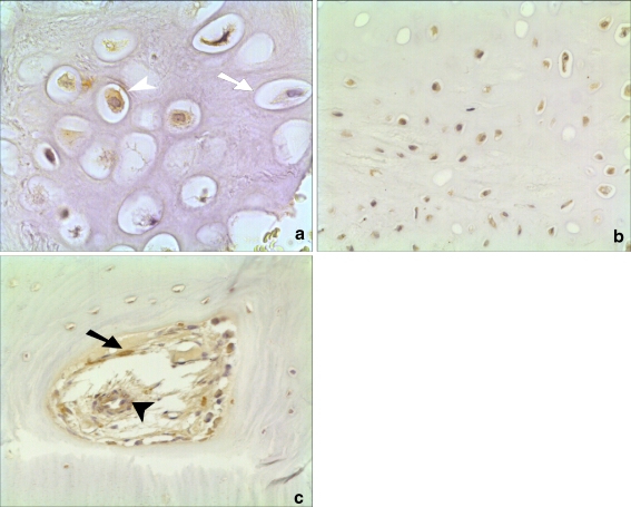
Fig. 1.
Histological appearance of noggin expression in a healing human fracture. a High-powered view (×40 magnification) showing brown staining in the nucleus of a hypertrophic chondrocyte (open arrowhead) compared to a non-stained cell (open arrow). b Staining in both hypertrophic and non-hypertrophic chondrocytes (×20). c Staining in new blood vessel forming in human fracture callus (closed arrowhead) and in osteoblast (closed arrow) (×20)

