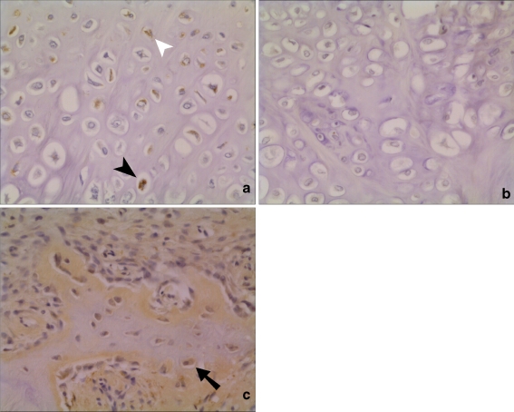
Fig. 2.
Histological appearance of chordin expression in human fractures (×20 view). a and b Serial sections of an area of cartilage formation. Staining in both hypertrophic (closed arrowhead) and non-hypertrophic chondrocytes (open arrowhead), with chordin antibody (a). No staining in when negative control IgG antibody was used as primary antibody during immunohistochemistry (b). c Expression of chordin in osteoblasts in area of active bone formation

