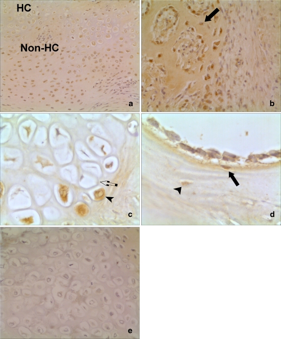
Fig. 4.
Histological appearance of BMP-2 expression in human fracture calluses. a and c Pattern of expression in area of cartilage formation. Staining predominant in non-hypertrophic chondrocytes (non-HC) compared to hypertrophic chondrocytes (HC) seen in a (×10). c Large power view (×40) of cytoplasmic staining in chondrocyte (arrowhead) compared to a non-staining cell adjacent (arrow). b (×10) and d (×40) Expression of BMP-2 in area of bone formation. Osteoblasts demonstrating BMP-2 expression are demonstrated with an arrow. There was weak expression of BMP-2 in osteocytes (arrowhead). e (×20) No staining when negative control IgG antibody used as primary antibody during immunohistochemistry. This section is a serial section of the one in (a)

