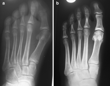
Fig. 3.
a Preoperative anteroposterior weight bearing radiograghs of left foot of 16-year-old female shows the obliquity of the first metatarsomedial-cuneiform joint. b Twelve years after the open wedge osteotomy of the cuneiform, the radiograph shows the decrease of medial inclination of this joint

