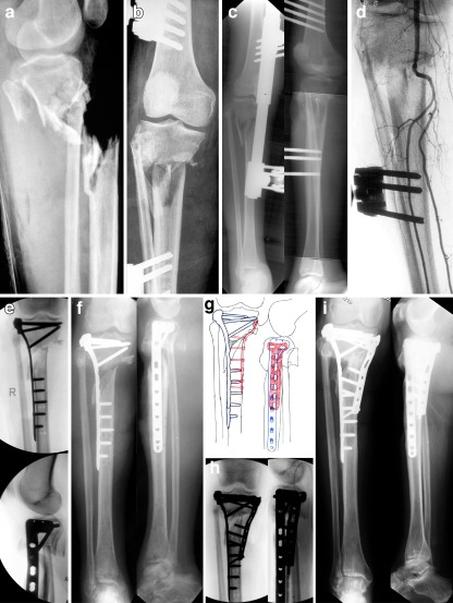
Fig. 2.
A 65-year-old female was skiing and was struck by another skier who was airborne after going off of a jump. She sustained a right-sided Grade IIIC open proximal tibia fracture with segmental metaphyseal bone loss. She was taken to a local trauma centre and also had extensive degloving. A joint spanning external fixator was applied and a vascular reconstruction was performed bypassing her popliteal trunk. She was transferred to our institution at three months following injury for definitive management of her tibia fracture. Presence of an eschar was noted near the fracture site and she had a viable foot but did have a foot drop with numbness on the dorsum and distal plantar aspect of her foot. She underwent multiple irrigation and debridements followed by removal of the external fixator, open reduction and internal fixation (ORIF), correction of varus deformity, and placement of a proximal tibia locking plate. The majority of the anterior compartment was debrided and a free rectus flap was performed to cover the proximal aspect of the tibia followed by a split thickness skin graft for skin coverage. She continued progress but at six months following ORIF, radiographs revealed a nonunion with maintenance of reduction and hardware. Revision ORIF was planned and performed with flap and muscle elevation, placement of DBM bone graft, BMP-7 and a medial proximal tibia locking plate for additional stability. She returned for regular follow-up intervals and at 39 months she presented with excellent radiographic and clinical results including a healed proximal tibia nonunion in excellent alignment with presence of partial lateral defect, significant improvement in pain, recovery of foot function and resolution in numbness symptoms, and a return to all pre-injury activities including skiing. Anteroposterior (AP) (a) and lateral (b) injury radiographs revealing Grade IIIC open proximal tibia fracture with segmental metaphyseal bone loss. c Radiographs following application of a spanning external fixator. d Arteriogram three months following popiteal truck bypass procedure. e Intraoperative fluoroscopic images following ORIF, correction of varus deformity and placement of a proximal tibia locking plate. f AP and lateral injury radiographs at five months revealing a proximal tibia nonunion with maintenance of reduction and hardware. g Preoperative plan for revision ORIF. h Intraoperative fluoroscopic images following ORIF, placement of DBM, BMP-7 and a medial proximal tibia locking plate. i AP and lateral radiographs at 39 months revealing a healed proximal tibia nonunion in excellent alignment with presence of partial lateral defect

