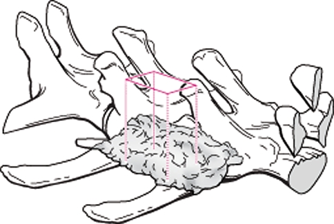
Fig. 1.
The bone mineral density was assessed using a Lunar DPXL DEXA machine. A region of interest within the center of the implant encompassing the entire fusion mass (excluding the transverse processes) on the right and left sides was used. DEXA density (g/cm2) increased with time for all FormaGraft groups, while the autograft decreased with time

