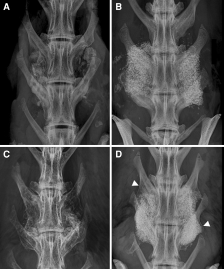
Fig. 2.
Typical radiographs are presented for autograft at 6 and 12 weeks (a and c) and FormaGraft alone at 6 and 12 weeks (b and d). The increase in fusion maturity with autograft as well as FormaGraft is present with the development of neocortex laterally. A progression of new bone enveloping the graft emanating from the transverse processes (arrows) was found between 6 and 12 weeks, which was also accompanied by resorption of the implant material in that area

