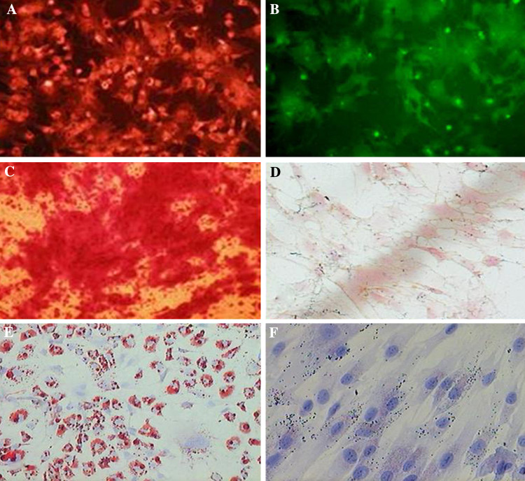
Fig. 1.
MSC surface marker expression was determined by immunocytochemically using the monoclonal antibodies (a) CD29-PE and (b) CD44-FITC as markers. Localized perinuclear pattern was observed (magnification, ×200). c MSCs were cultured for 2 weeks in osteogenic medium and stained with alizarin red S stain to identify differentiated cells. Several red stained regions, indicative of the presence of a calcified extracellular matrix, were observed (magnification, ×200). d No calcification was observed in undifferentiated MSCs maintained in control osteogenic medium (magnification, ×200). e The MSCs were cultured for 2 weeks in the adipogenic medium and stained with Oil Red O. A significant fraction of the cells contained many intracellular lipid-filled droplets that had accumulated Oil Red O (magnification, ×200). f No lipid droplets were observed in undifferentiated MSCs maintained in control adipogenic medium (magnification, ×200)

