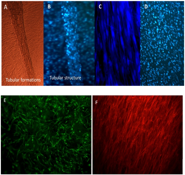
Figure 6.
C2C12 cells observed by phase contrast microscopy (A) shows formation of tubular structure from day 6th of culture. On further analysis by fluorescent microscopy using DAPI multinucleated tubular structure was seen to be formed by group of cells (B) along with background of cells spreading homogenously on the HG matrix. On 2-D culture the 6th day cells are seen after Hoechst staining (C) and DAPI staining (D). Cell tracker showing the proliferation of cells on the scaffold (E), and the alignment of the C2C12 in preferential direction on the scaffolds (F).

