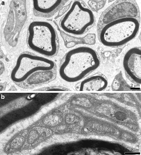
Fig. 2.
Transmission electron microscopy (TEM) control specimen. a Myelin sheats of Schwann cells enveloped the axons. Nuclei and intracellular organelles were intact. Scale bar 2 μm. b Unmyelinated fibre. Subtle sheat of single Schwann cells membrane enveloped numerous bundles of axons. Scale bar 2 μm

