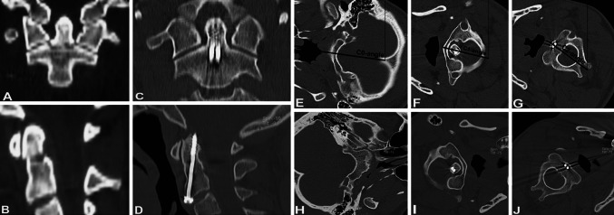
Fig. 2.
Patient with non-displaced odontoid type III fracture. Injury CT-scans depict transverse fracture course both in coronar and sagittal plane and treatment was with double AOSF. Eight years follow-up revealed solid union. Dynamic CT-scan in right head rotation showed C0-angle of 82.5°, C1-angle of 78.8° and C2-angle of 51.4° resembling an atlantoaxial separation angle of 27.4°. Accordingly, C1–2 rotation accounted for 33.2% of total head rotation. Patient had no malalignment and excellent self-rated clinical outcome. The atlantoaxial joints were unremarkable and congruent

