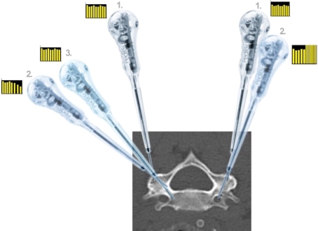
Fig. 2.
Principles in using an ECD for cervical pedicle tract preparation (Here: pCPS in C5). Right pedicle Following entry point selection and cortex perforation with the ECD (position 1), the ECD is inclined medially in direction of the anatomical pedicle axis (positions 1–2). If there is, e.g., to much a medial inclination the tip of the ECD will face cortical bone. Thus, while progressing the ECD towards the cortical wall, the ECD will emit signals with decreased frequency and sounds signaling that the ECD is at a cortex (position 2). Accordingly, as illustrated, the ECD is only 14 mm inside the vertebra and therefore the surgeon has to redirect the ECD (positions 2–3). After redirection the ECD will protrude again in cancellous bone and the ECD will emit homogenous signals indicating correct pedicle protrusion. Left pedicle Example of perforation of the transverse foramen. As on the right side, the ECD is driven into the lateral mass cancellous bone (position 1). If surgeon does not address the steep medial inclination of the anatomical pedicle axis, but rather goes straightforward he will breach the transverse foramen with the ECD (position 2). However, if this is done slowly, surgeon will face sharp signal changes emitted by the ECD with high pitching and highly frequenced signals indicating redirection of the ECD or abortion of pCPS fixation and change to lateral mass placement for that vertebra. The same principle is with ATPS insertion. The main difference is the larger corridor towards the pedicle and working area

