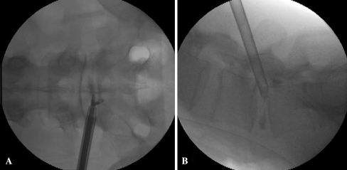
Fig. 1.
a Fluoroscopic images showing the position of the tip of working cannula in anteroposterior view. See the endoscopic forceps grasping the discal cyst in downward direction. b Fluoroscopic images showing the position of the tip of working cannula anchored in the subannulus in the lateral view, and the slight downward inclination of the needle trajectory on lateral view

