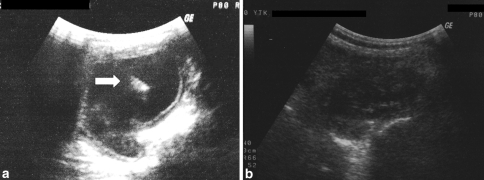
Fig. 1.
a Ultrasonographic appearance of the cyst at initial admission. Arrow shows the detached germinative membrane within the cyst indicating Type II hydatid cyst according to Gharbi classification. b Twenty-six months after the PAIR treatment, cyst showed decrease in dimensions, solidification of the content and thickening of the cyst wall

