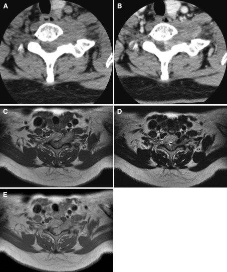
Fig. 1.
a Non-enhanced axial CT demonstrates a well-defined mass with neural foraminal widening at C6–7. The mass shows similar homogenous density, relative to the skeletal muscle. b Contrast-enhanced axial CT shows heterogeneous, well-enhanced mass at same levels. c Axial T1-weighted image shows a dumbbell-shaped, homogeneous mass with slight hyperintensity, compared with the spinal cord at the C6–7 level. d Axial T2-weighed MR images show a slightly hypointense mass with a dark signal portion at the anterolateral aspect of the mass. e Gadolinium-enhanced T1-weighted image show mild heterogeneous enhancement of the mass

