Abstract
Diffuse idiopathic skeletal hyperostosis (DISH) is a common systemic disorder characterised by the ossification of the anterior longitudinal spinal ligament involving at least three contiguous vertebrae and by diffuse extraspinal enthesopathies. The condition is associated with the male sex and with advanced age; its aetiology is uncertain, but seems to be related to obesity and diabetes. The most recent studies in archaeological series demonstrated a relation between high social status and the incidence of DISH. The present study examines two cases of DISH found amongst the members of the Medici family buried in the Basilica of San Lorenzo in Florence. The skeletons of the Grand Dukes Cosimo I (1519–1574) and his son Ferdinand I (1549–1609) showed the typical features of the condition. This result is related to the obesity of the Grand Dukes, attested by the written and artistic sources, and to the protein-based alimentation demonstrated by a paleonutritional study, thus furnishing further evidence to the significance of DISH as a life style.
Keywords: DISH, Morbus Forrestier, Spinal ankylosis, Obesity, Diabetes, Life style, Renaissance
Introduction
Diffuse idiopathic skeletal hyperostosis (DISH) is a common systemic disorder, first identified in 1950 by Forestier and Rotes-Querol [5], whose main characteristic is the ossification of the anterior longitudinal spinal ligament, with large osteophytes that flow down the spine, producing a typical candle wax appearance. These lesions occur most commonly in the thoracic portion of the column, although they may involve all the spinal segments. In the thoracic spine, the limitation of the changes to the right side is attributed to the presence of the left-sided descending aorta, which would prevent ossification. The fusion of at least four contiguous vertebrae is necessary for the diagnosis of DISH, but some authors estimate that three vertebrae are sufficient [16]. Ossification leads to ankylosis of the involved vertebrae, but intervertebral disc spaces and apophyseal joints are preserved [1]. DISH is known to be commonly associated with the ossification of other ligaments of the vertebral column [4] and of the ligament and tendon attachments of the appendicular skeleton [13, 15]. Osteoarthritis is often associated with DISH. In most modern clinical investigations, DISH is rarely observed in subjects under the fourth decade of life and its incidence progressively increases with advancing age; the condition shows predilection for the male sex [22]. The aetiology of DISH has been related to metabolic disorders, in particular obesity and type II diabetes mellitus [3, 18].
Methods
The “Medici Project”, a multidisciplinary research aimed to perform an archaeological and palaeopathological study on the 49 funerary depositions of the Medici Grand Dukes in the famous Medici Chapels of the Church of San Lorenzo in Florence, was a unique opportunity to reconstruct the health and life style of the members of this important Italian Renaissance family. The Medici were patrons of several of the great artists of the period, promoting the arts and sciences. Up till now, a total of 15 tombs have been explored, including those of the Grand Dukes Cosimo I (1519–1574) and Ferdinand I (1549–1609) [8, 9].
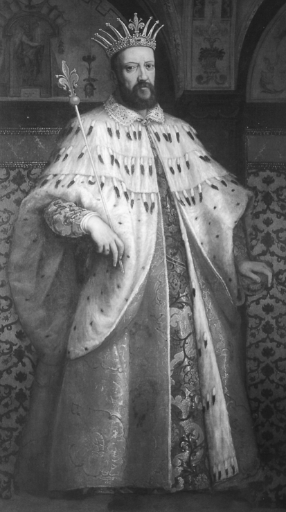
Cosimo I (Fig. 1) was son of Giovanni delle Bande Nere (1498–1526) and Maria Salviati (1499–1543). He became Duke of Florence in 1537 and was named first Grand Duke of Tuscany in 1569, governing until his death. In 1539, he married Eleonora from Toledo (1519–1562). They had 11 children.
Fig. 1.
Official portrait of Cosimo I revealing severe obesity (Cigoli, Florence, Medici-Riccardi Palace)
Ferdinand I (Fig. 2) was the sixth male son of Cosimo I and Eleonora from Toledo. In 1587, he became the third Grand Duke of Tuscany, ruling until his death in 1609. In 1589, he married Christine from Lorraine and they had nine sons.
Fig. 2.
Official portrait of Ferdinand I, revealing evident obesity (Tiberio Titi, Pisa, S. Matteo Museum)
After exhumation, the two skeletons were studied in a temporary laboratory set-up on the site and submitted to macroscopic and radiological examination.
Results
Cosimo I: Cosimo underwent a series of changes located along both the axial and appendicular skeleton.
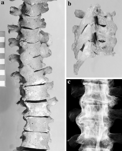
In the column, the anterior right-sided longitudinal ligament was ossified at the level of the T6, T7 and T8 vertebral bodies, forming a bony bridge between the vertebrae, which appeared as a continuous line of bumps (Fig. 3a–c). The intervertebral disks and the articular surfaces were preserved. Syndesmophytes, without vertebral fusion, were also present on other thoracic and lumbar vertebrae. Other two vertebrae, L2 and L3, were fused through a bony bridge on the left side. Marked bone spurs at the insertion of the ligamenta flava insertion were also observed. Enthesopathies were present in several ligament and tendon attachments. The skeleton of Cosimo showed diffuse and severe arthritis, affecting the joints of the axial and appendicular skeleton.
Fig. 3.
The DISH of Cosimo I: a anterior view of the column with ossification of the anterior right vertebral ligament, at the level of the 6th–8th thoracic vertebral bodies, b right lateral view and c anteroposterior X-ray of the T6–T8 block
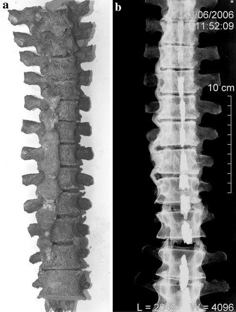
Ferdinand I: The vertebral bodies from T5 to T9 were fused in a unique block owing to the ossification of the right anterior ligament, which conferred the typical aspect of a “candle wax” to this spine segment (Fig. 4a, b). Another left-sided fusion was observed at the level of the T10 and T11 bodies. Ossification of the right anterior ligament with no formation of bony bridges, was observed in other cervical and thoracic vertebrae. Intervertebral spaces and apophyseal joints were normal. Several vertebral ligament insertions, as well as extraspinal ligaments, showed large ossifications. Ferdinand suffered from diffuse osteoarthritis (involving the spine as well as the great and small joints of the appendicular skeleton) and from chronic left foot gout, also diagnosed at palaeopathological study [7].
Fig. 4.
The DISH of Ferdinand I: a anterior view of the column with ossification of the anterior right vertebral ligament, at the level of the 5th–11th thoracic vertebral bodies, conferring the typical “candle wax” appearance, b Anteroposterior X-ray of the thoracic column
Discussion
The changes observed in Cosimo I and Ferdinand I meet the standard major criteria established for the diagnosis of DISH [17]. Both skeletons show ossification of the right anterior ligament of the column with the involvement of at least three vertebrae in Cosimo and, in the case of Ferdinand, even of seven vertebrae, and also diffuse ossification of many articular ligaments and entheses.
A number of palaeopathological cases have been attested in various geographical sites and at different periods, whereas extensive studies have evaluated the incidence of DISH in large skeletal series (see references in [17]). Another case in the Medici family has to be added to those found by us, that of Cosimo “the Elder” (1389–1464), whose remains were exhumed and studied during the Second World War. The macroscopic and radiological examinations showed the ossification of the anterior longitudinal ligament from T6 to T12 included [2].
Following the clinical studies that demonstrated an association between DISH and obesity, diabetes and hyperinsulinemia [19], a recent paleopathological investigation has related the incidence of DISH with the social status and life style, with particular regard to nutritional patterns [11]. A study conducted at Merton Priory (Surrey, UK) showed a DISH prevalence of 8.6% on 35 monkish burials, which is considerably higher than the figures found in other large archaeological series or modern populations. In this study, Waldron [21] was the first to relate the results to the monastic dietary habits, characterised by the consumption of a rich and high-calorie diet, causing possible obesity and late-onset diabetes. As a matter of fact, medieval documentation describes an alimentation based on the animal fats and alcoholic drinks, with average high-calorie intake.
Further investigations support the observations made by Waldron. In the cemetery of Wells Cathedral and Royal Mint in London a statistically significant difference was observed between the priests’ and lay-benefactors’ burials in the churches and chapels, which showed higher values, and those of the general population in the “normal” cemeteries [17]. The authors explain this variation mainly due to occupational factors and differences in food access.
More evident are the results from the Basilica of Saint Servaas in Maastricht, where all 27 skeletons of the canons were affected by DISH [12]. Predisposition of the “monastic way of life” to DISH is further corroborated by the recent study on the presumable clergymen and high-ranking citizens excavated from the abbey court in Maastricht [20]. In this sample, 40.4% of the adult population showed features satisfactory for a diagnosis of DISH.
The Medici family represents an ideal case study for investigating the life-style patterns of the high social classes. The Italian Renaissance aristocratic classes had access to a wide variety of food. The historical archives describe a diet-based essentially on meat and wine, occasionally enriched with eggs and cheese and, on penitential occasions, with fish. The consumption of vegetables was scarce and fruit was almost totally absent from the alimentation [10].
The archival documents also refer that Cosimo was rather moderate with regard to alimentation. However, the pictorial sources show a well-built individual, and the portraits confirm that he became corpulent in the last decades of life (Fig. 1). Ferdinand was rather fat during all his life, and become obese after 40 years of age, as testified by the written and artistic sources (Fig. 2). The Venetian representative in Florence Giovanni Gritti states that the Grand Duke, who was aged 39 years, “is of wet and fat constitution, whence it is dubious that he will live longtime” [14].
A paleonutritional study, performed recently on the Medici Grand Dukes and their families, has confirmed the written sources. Carbon and nitrogen stable isotope analysis revealed a diet very rich in meat, as the δ15N high values at the level of carnivores demonstrate. The δ13C values, related to the consumption of fish, confirmed an intake of marine proteins at 14–30% [6].
Therefore, the association between DISH and elite status, already highlighted in recent investigations [11] receives further confirmation in the present study. Of the three Medici male individuals over 40 years of age studied so far, two were affected by DISH, as demonstrated by this study. Despite the narrowness of the sample, the high incidence of DISH in members of a high social class, like the Medici of Renaissance Florence, is noteworthy, having significance as a life-style marker and supporting the link between nutritional patterns and risk of developing DISH in mature age.
Acknowledgment
This study is integral part of the “Medici Project”, performed with the permission of the Superintendence for Florentine Museums.
Conflict of interest statement None of the authors has any potential conflict of interest.
References
- 1.Cammisa M, Serio A, Guglielmi G. Diffuse idiopathic skeletal hyperostosis. Eur J Radiol. 1998;27:S7–S11. doi: 10.1016/S0720-048X(98)00036-9. [DOI] [PubMed] [Google Scholar]
- 2.Costa A, Weber G. Le alterazioni morbose del sistema scheletrico in Cosimo dei Medici il Vecchio, in Piero il Gottoso, in Lorenzo il Magnifico, in Giuliano Duca di Nemours. Arch de Vecchi Anat Patol. 1955;23:1–69. [PubMed] [Google Scholar]
- 3.Denko CW, Malemud CJ. Body mass index and blood glucose: correlations with serum insulin, growth hormone, and insulin-like growth factor-1 levels in patients with diffuse idiopathic skeletal hyperostosis (DISH) Rheumatol Int. 2006;26:292–297. doi: 10.1007/s00296-005-0588-8. [DOI] [PubMed] [Google Scholar]
- 4.Ehara S, Shimamura T, Nakamura R, Yamazaki K. Paravertebral ligamentous ossification: DISH, OPLL and OLF. Eur J Radiol. 1998;27:196–205. doi: 10.1016/S0720-048X(97)00164-2. [DOI] [PubMed] [Google Scholar]
- 5.Forestier J, Rotes-Querol J. Senile ankylosing hyperostosis of the spine. Ann Rheum Dis. 1950;9:321–330. doi: 10.1136/ard.9.4.321. [DOI] [PMC free article] [PubMed] [Google Scholar]
- 6.Fornaciari G. Food and disease at the renaissance courts of naples and florence: a paleonutritional study. Appetite. 2008;51:10–14. doi: 10.1016/j.appet.2008.02.010. [DOI] [PubMed] [Google Scholar]
- 7.Fornaciari G, Giuffra V, Giusiani S, Fornaciari A, Villari N, Vitiello A (2009) The “Gout” of the Medici, Grand Dukes of Florence: a Palaeopathological Study. Rheumatology (in press) [DOI] [PubMed]
- 8.Fornaciari G, Vitiello A, Giusiani S, Giuffra V, Fornaciari A. The “Medici Project”: first results of the explorations of the Medici tombs in Florence (15th–18th centuries) Paleopathol News. 2006;133:15–22. [Google Scholar]
- 9.Fornaciari G, Vitiello A, Giusiani S, Giuffra V, Fornaciari A, Villari N. The “Medici Project”: first anthropological and paleopathological results of the exploration of the Medici Tombs in florence (15th–18th Centuries) Med Secoli. 2007;19(2):521–544. [PubMed] [Google Scholar]
- 10.Grieco AJ. Food and social classes in Late Medieval and Renaissance Italy. In: Flandrin JL, Montanari M, editors. Food: a culinary history from antiquity to the present. New York: Columbian University Press; 1999. pp. 302–312. [Google Scholar]
- 11.Jankauskas R. The incidence of diffuse idiopathic skeletal hyperostosis and social status correlations in Lithuanian skeletal materials. In J Osteoarchaeol. 2003;13:289–293. doi: 10.1002/oa.697. [DOI] [Google Scholar]
- 12.Janssen HAM, Maat GJR. Canons buried in the “Stiftskapel” of the Saint Servaas Basilica at Maastricht AD 1070–1521. A Palaeopathological Study. Leiden: Barge’s Anthropologica; 1999. [Google Scholar]
- 13.Littlejohn GO, Urowitz MB. Peripheral enthesopathy in diffuse idiopathic skeletal hyperostosis (DISH): a radiologic study. J Rheumatol. 1982;9:568–572. [PubMed] [Google Scholar]
- 14.Pieraccini G. La stirpe dei Medici di Cafaggiolo. Firenze: Nardini Editore; 1986. [Google Scholar]
- 15.Resnick D, Shaul S, Robins J. Diffuse idiopathic skeletal hyperostosis with extraspinal manifestations. Radiology. 1975;115:513–524. doi: 10.1148/15.3.513. [DOI] [PubMed] [Google Scholar]
- 16.Rogers J, Waldron T. A field guide to joint disease in archaeology. Chichester: Wiley; 1995. [Google Scholar]
- 17.Rogers J, Waldron T. DISH and the Monastic way of life. Int J Osteoarchaeol. 2001;11:357–365. doi: 10.1002/oa.574. [DOI] [Google Scholar]
- 18.Sarzi-Puttini P, Atzeni F. New developments in our understanding of DISH (diffuse idiopathic skeletal hyperostosis) Curr Opin Rheumatol. 2004;16:287–292. doi: 10.1097/00002281-200405000-00021. [DOI] [PubMed] [Google Scholar]
- 19.Smythe H, Littlejohn G (1998) Diffuse idiopathic skeletal hyperostosis. In: Klippel JH, Dieppe PA (eds) Rheumatology, 2nd edn, vol 2. Mosby, London, pp 8.10.1–6
- 20.Verlaan JJ, Oner FC, Maat GJR. Diffuse idiopathic skeletal hyperostosis in ancient Clergymen. Eur Spine J. 2007;16:1129–1135. doi: 10.1007/s00586-007-0342-x. [DOI] [PMC free article] [PubMed] [Google Scholar]
- 21.Waldron T. DISH at Merton Priory: evidence for a “new” occupational disease? Br Med J. 1985;291:1762–1763. doi: 10.1136/bmj.291.6511.1762. [DOI] [PMC free article] [PubMed] [Google Scholar]
- 22.Westerveld LA, Ufford HME, Verlaan JJ, Oner FC. The prevalence of diffuse idiopathic skeletal hyperostosis in an outpatient population in the Netherlands. J Rheum. 2008;35(8):1635–1638. [PubMed] [Google Scholar]


