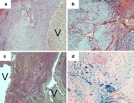
Fig. 2.
Cavernous vascular malformation, histopathological features: prominent venous vessel with an excessively thickened, amuscular wall (a, H&E). Hyalinized vessels (b, H&E) are arranged in a back-to-back fashion (c, Elastica van Gieson stain) as typical for cavernous angioma. Signs of recurrent hemorrhage with fresh bleedings (b) and residues of older bleedings in form of hemosiderin deposition (d, Berlin blue stain). Magnification of images a and c, ×100; magnification of images b and d, ×200; V vessel lumina

