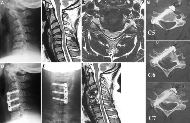
Fig. 4.
Imaging studies of the illustrative case. a Preoperative lateral radiograph showing C5 compression fracture and kyphotic deformity. b, c T2 sagittal and axial images of MR demonstrating spinal cord compression and obscure changes in cord signals. d, e Postoperative AP and lateral radiographs showing good alignment and C4–6 pedicle screw fixation. f Postoperative T2 sagittal image of MR showing good decompression and neutral alignment. g Postoperative axial CT demonstrating good placement of anterior pedicle screws at C4–6 levels

