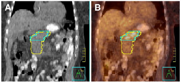
FIGURE 4.

Pancreatic adenocarcinoma delineated on CT alone by radiation oncologist (yellow) and on 18F-FDG PET/ CT by nuclear medicine physician (blue). Images are from coronal cut of patient with CT (A) and 18F-FDG PET/CT image fusion (B). Tick marks are 1 cm.
