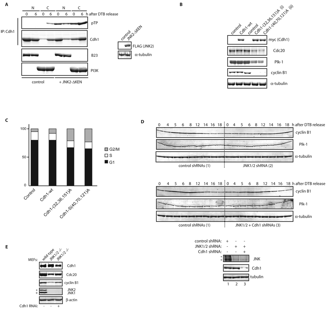
Figure 6. JNK-mediated phosphorylation of Cdh1 regulates its function.
(A) In vitro kinase assays using active JNK and MBP-Cdh1 wild-type (wt) or single phosphorylation sites mutants. (B) In vitro kinase assays using active JNK and Cdk1 either alone or sequentially (first reaction –priming– was performed using cold ATP) and recombinant MBP-Cdh1 as substrate. (C) Unphosphorylated or in vitro phosphorylated 6×His-Cdh1 by JNK, were used to pull-down Cdc27 from extracts produced from exponentially growing cell lines. (D) In vitro ubiquitination assay using APC/C complex immunoprecipitated from HeLa cells and either unphosphorylated or JNK-phosphorylated Cdh1 and 6×His-TOME-1 as substrate. Ubiquitination reactions were performed in the presence of 32P-labeled ubiquitin (previously phosphorylated by PKA in vitro) and were analyzed by SDS-PAGE and PhosphorImaging. Membrane was probed with TOME-1 antibodies to detect levels of unmodified substrate. (E) Nuclear (N) and Cytosolic (C) fractions produced from HeLa cells synchronized by a DTB and released (0, early S-phase arrest; and 6 h, G2 phase) into control or JNK VII inhibitor-containing media (JNKi; 10 µM added at 4 hours release time-point). Extracts prepared from these fractions were analyzed by Cdc27 immunoblotting and subjected to Cdh1 immunoprecipitation followed by Cdh1 and phospho-ThrPro (pTP) immunoblotting. B23/nucleophosmin and PI3K serve as nuclear and cytoplasmic markers, respectively. Levels of JunB (whose reduction is a marker of G2-phase) were assessed in the 0 h (double-thymidine blocked) versus 6 h-released (G2) extracts, only for the control conditions. FACS analyses are also included. Uncropped images for key results of this figure are shown in Figure S7.

