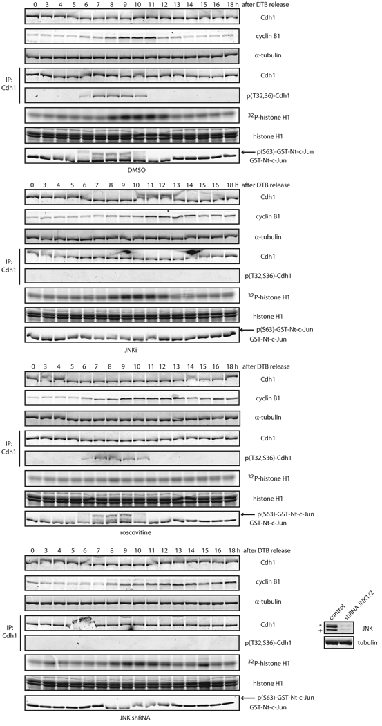
Figure 7. JNK phosphorylates Cdh1 in cells independently of CDKs activation.
Phosphorylation status of endogenous Cdh1 after immunoprecipitation at residues Threonine 32 (T32) and Serine 36 (S36) in cell cycle-synchronized HeLa cells after a double-thymidine block (DTB). JNKi (JNK VII inhibitor) was used at 10 µM at the 4 hours release time-point. Roscovitine was utilized at 100 µM at the 6 hours release time-point. Down-regulation of JNK1 and JNK2 achieved by means of shRNA, is shown in the inset panels (bottom right). Western-blot corresponds to the 0 h time-point; JNK2 displays as a 54kDa band (*) while JNK1 displays as a 46kDa band (+). CDKs assays were performed in vitro –using total extracts– by assessing 32P-γ-ATP incorporation in histone H1 as substrate. JNK activity was assessed in vitro, using total extracts incubated with cold ATP and recombinant GST-N-terminus-tagged c-Jun, and revealed by immunoblotting using phospho-Ser63-c-Jun antibodies following a GSH-pull down. Uncropped images for key results of this figure are shown in Figure S7.

