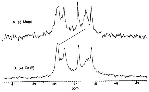
FIGURE 3.
Binding of Ca(II) to the D-glucose-occupied, 3F-Phe-labeled receptor: 19F NMR spectra. 3F-Phe incorporation was 20 ± 10%. Samples contained 200–500 μM labeled receptor, 100 mM KCl, 10 mM Tris, pH 7.1 with HCl, 1 mM D-glucose, 10% D2O, and (A) 20 mM EDTA or (B) 0.5 mM CaCl2. Spectral parameters at 470 MHz were as in Figure 1, except that 5F-Trp was used as an internal frequency standard.

