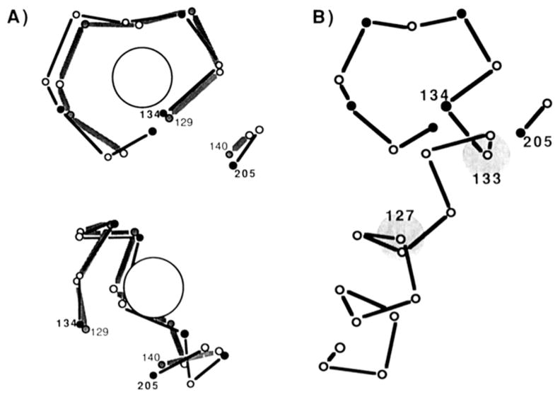
FIGURE 1.

α-Carbon backbone structures of two Ca(II) sites: cal-modulin site IV and the GGR site. (A) Two views of the corresponding backbone regions of calmodulin site IV [gray line, residues 129–136 and 139–140 (Babu et al., 1988)] and of the GGR site [bold line, residues 134–142 and 204–205 (Vyas et al., 1987)]. The position of bound Ca(II) in calmodulin site IV is indicated by the large circle. Ligand positions are indicated by filled vertices in the backbone. For clarity the corresponding regions of calmodulin site IV and GGR sites are slightly offset; also, the regions of divergence are omitted (calmodulin site IV residues 137–138 are omitted, and GGR site residues 142–203 are omitted). (B) A view of the GGR site and a portion of the preceding helix (Vyas et al., 1987). The circles at helix positions 127 and 133 indicate the backbone positions of nearby tryptophan residues.
