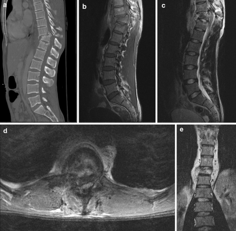
Fig. 5.
A 28-year-old man was injured in a motor vehicle accident. CT scan of the thoracolumbar spine was performed with sagittal mutiplanar reformations (a). There is an unstable burst fracture of Th11 with retropulsion of bone fragments into the spinal canal, and kyphotic angulation. In order to assess the spinal cord, MRI of the thoracolumbar spine was performed with sagittal T1-weighted images (b), sagittal T2-weighted images (c), axial T2-weighted images (d). The spinal canal is narrowed with extrinsic compression of the spinal cord and intramedullary focal areas of hyperintensity, indicating spinal cord oedema. The coronal T1-weighted image (e) shows the paravertebral hematoma

