
Fig. 4.
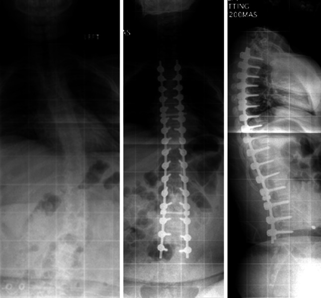
Plain radiographs showing pre-operative and post-operative radiographs in a DMD patient with scoliosis. Pedicle screw instrumentation has been used to achieve a good correction in this case. Notice the placement of pedicle screws at all levels and that instrumentation has been restricted to L5