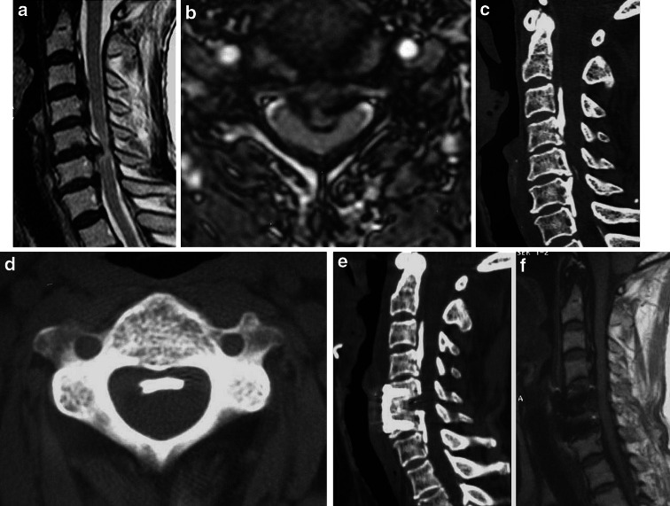
Fig. 3.
A 67-year-old patient with spinal cord compression mainly from C5/6 disc hernia but not from OPLL. a, b Sagittal and axial MRI scanograms of the cervical spine showing Grade II compression from OPLL, and the maximum compression at C5/6 disc level. c Sagittal 3D CT showing mixed-type OPLL from C3 to C7. d Axial scanogram showing a flat ossified ligament. e Sagittal 3D CT after ACDF and instrumentation showing an adequate decompression. f Sagittal MRI showing good spinal cord appearance after ACDF

