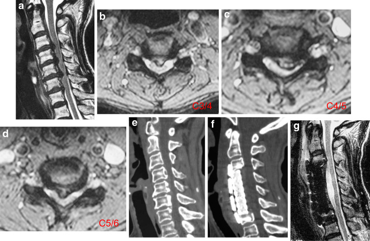
Fig. 4.
A 57-year-old patient with spinal cord compression from disc hernia, and OPLL with no more than three segments and a 50% occupying rate. a Sagittal MRI of the cervical spine showing Grade III compression due to OPLL and the maximum compression at disc level. b–d Axial scanograms showing C3/4, C4/5, and C5/6 disc hernias. e Sagittal 3D CT showing segmental-type OPLL from C4 to C6. f, g Anterior corpectomy with fusion was performed and a good cord appearance was visible

