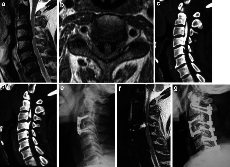
Fig. 5.
Spinal cord compression from anterior CDH and OPLL with occupying rate more than 50%. a, b Sagittal and axial MRI scanogram showing compression from OPLL, particularly at the C3/4 disc level. c, d Sagittal 3D CT showing continuous-type OPLL from C2 to C4, but an unossified area in the middle part of the longitudinal ligament at C3/4. e One-stage ACDF was performed. f Compression from OPLL at C3 and C4 remained. g Two-stage posterior laminectomy was performed

