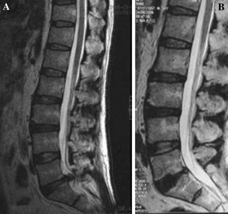
Fig. 6.
a Sagittal T2-weighted magnetic resonance image of the lumbar spine, showing degenerated, collapsed, and herniated disc at L5/S1 of a 52-year-old man with low back pain of 10 year duration with compressive radicular pain 2 year. b Follow-up radiograph 2 years postoperatively showing implanted B-Twin ESS. Disc height is partially restored

