
Fig. 1.
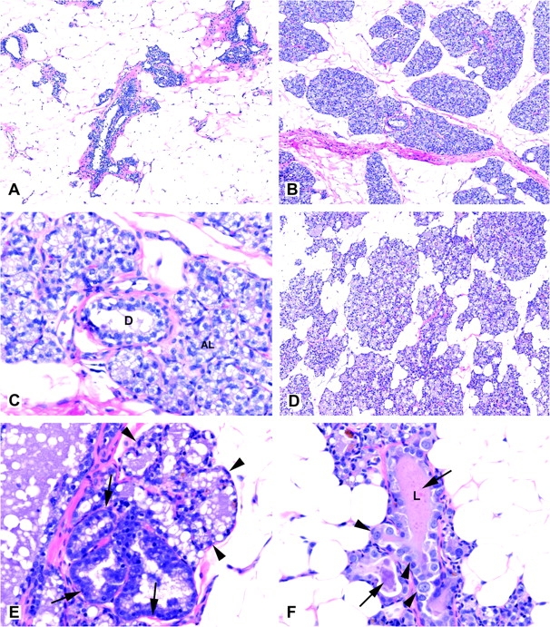
Histomorphological changes in mammary gland of female ACI rats exposed to 17β-estradiol for 6 and 12 weeks. (A) Mammary gland from untreated (control) ACI female rats (original magnification ×100); (B) Typical alveolar hyperplasia after 6 weeks of continuous E2 treatment (original magnification ×100); (C) Typical hyperplasia showing alveolar lobule (AL) and a normal appearing intralobular duct (D) after 6 weeks of continuous E2 treatment (original magnification ×400); (D) Typical hyperplasia after 12 weeks of continuous E2 treatment (original magnification ×100; (E) Mammary gland hyperplasia with atypical (arrows) and typical (arrowheads) hyperplastic alveoli after 12 weeks of continuous E2 treatment (original magnification ×100) and (F) Mammary gland hyperplasia with atypical hyperplastic ducts (arrows) line with variably sized epithelial cells (arrowheads). Duct lumen (L) contains intensely eosinophilic secretion; compare with normal appearing duct in panel C. Original magnification ×400.