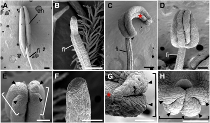
Figure 3.
Scanning Electron Microscopy Images of the Stamen in the Wild Type and rol.
(A) and (E) Wild type.
(B) and (F) Pin-like stamen.
(C) and (G) OT-type stamen with only one theca.
(D) and (H) ATT-type stamen showing four pollen sacs localized adaxially.
(E) to (H) Close-up views of (A) to (D), respectively.
Arrowheads and stars indicate the pollen sac and the connective, respectively. Brackets indicate a theca. an, anther; fi, filament. Bars = 200 μm in (A) to (D) and 100 μm in (E) to (H).
[See online article for color version of this figure.]

