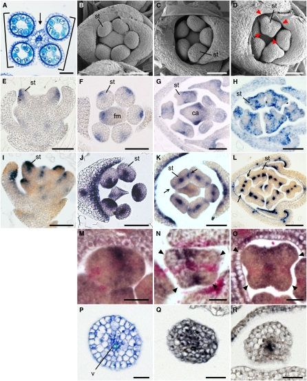
Figure 4.
Spatial Expression Pattern of ETT1 and PHB3 during Stamen Development in the Wild Type.
(A) Cross-section of an anther. Arrow and bracket indicate the connective and the thecae, respectively.
(B) to (D) Spikelets at the early developmental stages in the wild type. Arrowheads indicate protrusions in the stamen primordia.
(E) to (H) Spatial expression patterns of ETT1 in a longitudinal section (E) and cross sections ([F] to [H]) of the spikelet.
(I) to (L) Spatial expression patterns of PHB3 in a longitudinal section (I) and cross sections ([J] to [L]) of the spikelet. Arrows indicate the PHB3 expression domain in the lateral region of the stamen primordium.
(M) to (O) Two-color in situ hybridization in the anther. Purple, ETT1; pink, PHB3. Arrowheads indicate protrusions in the stamen primordia ([N] and [O]).
(P) Cross section of a filament.
(Q) and (R) Expression of ETT1 (Q) and PHB3 (R) in the filament.
ca, carpel; fm, floral meristem; st, stamen; v, vascular tissue. Bars = 50 μm in (A) to (L) and 20 μm in (M) to (R).

