
Fig. 5.
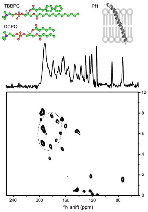
One-dimensional 15N chemical shift (centre) and two-dimensional 15N chemical shift/1H–15N dipolar coupling SLF (bottom) solid-state NMR spectra of uniformly 15N labelled Pf1 coat protein aligned with its bilayer normal parallel to the applied magnetic field in TBBPC:DCPC bicelles (X = 89% (q = 8), h = 80%, 40 °C). Top: structures of TBBPC and DCPC together with a sketch representing the helices’ orientations as deducted from spectra. (Adapted from [55]).