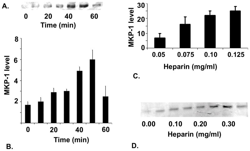
Figure 1. Heparin effects on MKP-1 levels in VSMCs.
Porcine VSMCs were grown to 70% confluence and starved for 48 hr. After starvation, cells were incubated with 100 μg/ml heparin (panel A) for various times. Cells were harvested as described in Methods and MKP-1 levels determined by Western blot analysis using anti-MKP-1 antibody. Panel A shows a representative blot of data from the five experiments included in the graph plotted below indicating increased MKP-1 levels over time of heparin treatment (panel B). Panel C, illustrates effects of heparin on porcine VSMCs over a heparin concentration range. Panel D illustrates the results when starved A7r5 cells were treated for 10 min with heparin at a wide range of concentrations.

