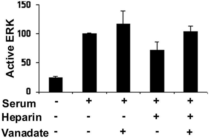
Figure 5. Vanadate intereferes with heparin effects on MAPK activity.

A7r5 cells were grown to approximately 60% confluence, starved and treated with 100 μg/ml heparin, 100 μM sodium vanadate, and activated with PMA for 15 min or left untreated as described in Methods. Active ERK was determined by Western blotting as described in the Methods. The absorbance determined for the PMA samples in each experiment was set to 100% and the other values were calculated as a percent of the PMA only samples to facilitate compiling the data from four experiments. The differences between PMA and PMA with vanadate and PMA with heparin and vanadate are not significant. All three are significantly different from PMA and heparin (p<0.05 for each case).
