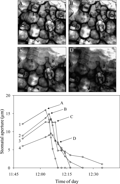
Figure 1.
Stomatal response in an isolated epidermis of T. pallida grafted to an exposed mesophyll of T. pallida after distilled water was added to the edge of the epidermis. Water spread between the mesophyll and epidermis by capillary action over the course of several minutes. Areas with water between the epidermis and mesophyll can be identified by the difference in contrast associated with the loss of air-water interfaces. Water is first visible in the top left corner of image A and spreads from left to right through subsequent images. White circles represent individual stomata on a single grafted epidermis and correspond to numbers in pictures; lettered arrows correspond to images. The experiment was repeated five times with similar results.

