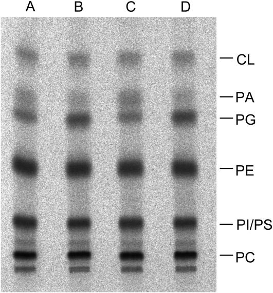
Figure 10.
In vivo [33P]phosphate labeling pattern of phosphoglycerolipids of the cds4 cds5 mutant and the wild type. After an 18-h incubation with labeled phosphate, lipids were extracted from the mutant (lanes A and C) and wild-type plants (B and D) and separated by thin-layer chromatography, and radioactively labeled lipids were visualized with a Bioimager and identified by cochromatography with authentic lipids (PC, phosphatidylcholine; PE, phosphatidylethanolamine; PI, phosphatidylinositol; PS, phosphatidylserine).

