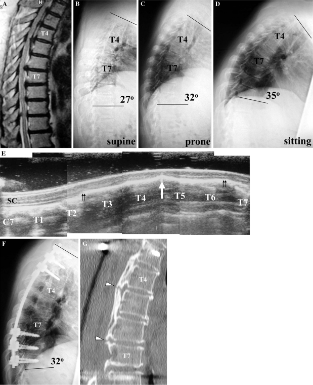
Fig. 1.
Preoperative T2-weighted MR image at the midsagittal plane (a) of a 55-year-old woman (Case 20), showing severe narrowing of the spinal cord at T4/5 and T6/7. Preoperative radiographic images show that the kyphotic angle at T1–T10 was 27° in the supine position (b), 32° in the prone position (c) and 35° in the sitting position (d). Intraoperative spinal ultrasonography at the midsagittal plane after laminectomy shows anterior impingement of the spinal cord by a beak-type OPLL at T4/5 (e, arrow) and absence of the subarachnoid space on the ventral side of the spinal cord from the T2/3 to T6/7 levels (e, double arrows). SC spinal cord. A postoperative radiographic image (f) shows a kyphotic angle at T1–T10 of 32°. A midsagittal reconstruction CT image (g) shows a non-ossified area at the mid-portion of the ossified mass at T4/5 and T6/7 (g, arrowheads)

