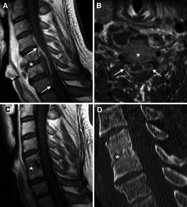
Fig. 3.
Preoperative imaging of a cervical spondylodiscitis with prevertebral und epidural abscesses, after surgical decompression and 9-months CT-control after monosegmental PEEK-cage spondylodesis. a, b Sagittal and axial gadolinium-enhanced T1-MRI imaging illustrates the spondylodiscitis mainly in segment C5/6 (star) with prevertebral (black arrow) and epidural abscesses (white arrows). c Gadolinium-enhanced T1-MRI imaging after 6 months demonstrates a completely decompressed spinal canal without any sign of a local infection. d Sagital CT images demonstrate the complete bony fusion of the involved level C5/6 1 year after monosegmental PEEK-cage spondylodesis. The stars mark the involved level C5/6. Again, there were no signs of neck pain, micro- or macroinstability in this patient

